This is default featured slide 1 title

Go to Blogger edit html and find these sentences.Now replace these sentences with your own descriptions.This theme is Bloggerized by Lasantha Bandara - Premiumbloggertemplates.com.

This is default featured slide 2 title

Go to Blogger edit html and find these sentences.Now replace these sentences with your own descriptions.This theme is Bloggerized by Lasantha Bandara - Premiumbloggertemplates.com.

This is default featured slide 3 title

Go to Blogger edit html and find these sentences.Now replace these sentences with your own descriptions.This theme is Bloggerized by Lasantha Bandara - Premiumbloggertemplates.com.

This is default featured slide 4 title

Go to Blogger edit html and find these sentences.Now replace these sentences with your own descriptions.This theme is Bloggerized by Lasantha Bandara - Premiumbloggertemplates.com.

This is default featured slide 5 title

Go to Blogger edit html and find these sentences.Now replace these sentences with your own descriptions.This theme is Bloggerized by Lasantha Bandara - Premiumbloggertemplates.com.

Wednesday, June 26, 2013

Antibiotic resistance


Antibiotic resistance


Antibiotic resistance tests: The bacteria in the culture on the left are sensitive to the antibiotics contained in the white, paper discs. The bacteria in the culture on the right are resistant to most of the antibiotics


Antibiotic resistance is a type of drug resistance that occurred by microorganism. Usually a bacterial species are able to survive against the exposure to one or more antibiotics; pathogens resistant to multiple antibiotics are considered multidrug resistant (MDR) or, more colloquially, superbugs. Microbes, rather than people, develop resistance to antibiotics.

Antibiotic resistance is a serious and growing phenomenon in contemporary medicine and has emerged as one of the pre-eminent public health concerns of the 21st century, particularly as it pertains to pathogenic organisms (the term is especially relevant to organisms which cause disease in humans). In the simplest cases, drug-resistant organisms may have acquired resistance to first-line antibiotics, thereby necessitating the use of second-line agents. Typically, a first-line agent is selected on the basis of several factors including safety, availability and cost; a second-line agent is usually broader in spectrum, has a less favourable risk-benefit profile and is more expensive or, in dire circumstances, be locally unavailable. In the case of some MDR pathogens, resistance to second and even third-line antibiotics is thus sequentially acquired, a case quintessentially illustrated by Staphylococcus aureus in some nosocomial settings. Some pathogens, such as Pseudomonas aeruginosa, also possess a high level of intrinsic resistance.

It may take the form of a spontaneous or induced genetic mutation, or the acquisition of resistance genes from other bacterial species by horizontal gene transfer via conjugation, transduction, or transformation. Many antibiotic resistance genes reside on transmissible plasmids, facilitating their transfer. Exposure to an antibiotic naturally selects for the survival of the organisms with the genes for resistance. In this way, a gene for antibiotic resistance may readily spread through an ecosystem of bacteria. Antibiotic-resistance plasmids frequently contain genes conferring resistance to several different antibiotics.

This is not the case for Mycobacterium tuberculosis, the bacteria that causes Tuberculosis, since evidence is lacking for whether these bacteria have plasmids. Also. M. tuberculosis lack the opportunity to interact with other bacteria in order to share plasmids.

Genes for resistance to antibiotics, like the antibiotics themselves, are ancient. However, the increasing prevalence of antibiotic-resistant bacterial infections seen in clinical practice stems from antibiotic use both within human medicine and veterinary medicine. Any use of antibiotics can increase selective pressure in a population of bacteria to allow the resistant bacteria to thrive and the susceptible bacteria to die off. As resistance towards antibiotics becomes more common, a greater need for alternative treatments arises. However, despite a push for new antibiotic therapies there has been a continued decline in the number of newly approved drugs. Antibiotic resistance therefore poses a significant problem.

The growing prevalence and incidence of infections due to MDR pathogens is epitomised by the increasing number of familiar acronyms used to describe the causative agent and sometimes the infection generally; of these, MRSA is probably the most well-known, but others including VISA (vancomycin-intermediate S. aureus), VRSA (vancomycin-resistant S. aureus), ESBL (Extended spectrum beta-lactamase), VRE (Vancomycin-resistant Enterococcus) and MRAB (Multidrug-resistant A. baumannii) are prominent examples. Nosocomial infections overwhelmingly dominate cases where MDR pathogens are implicated, but multidrug-resistant infections are also becoming increasingly common in the community.


Causes Antibiotic resistance:

Although there were low levels of preexisting antibiotic-resistant bacteria before the widespread use of antibiotics, evolutionary pressure from their use has played a role in the development of multidrug resistance varieties and the spread of resistance between bacterial species. In medicine, the major problem of the emergence of resistant bacteria is due to misuse and overuse of antibiotics. In some countries, antibiotics are sold over the counter without a prescription, which also leads to the creation of resistant strains. Other practices contributing towards resistance include the addition of antibiotics to livestock feed.

Household use of antibacterials in soaps and other products, although not clearly contributing to resistance, is also discouraged (as not being effective at infection control). Unsound practices in the pharmaceutical manufacturing industry can also contribute towards the likelihood of creating antibiotic-resistant strains.
The procedures and clinical practice during the period of drug treatment are frequently flawed — usually no steps are taken to isolate the patient to prevent re-infection or infection by a new pathogen, negating the goal of complete destruction by the end of the course.

Certain antibiotic classes are highly associated with colonisation with "superbugs" compared to other antibiotic classes. A superbug, also called multiresistant, is a bacterium that carries several resistance genes.  The risk for colonisation increases if there is a lack of sensitivity (resistance) of the superbugs to the antibiotic used and high tissue penetration, as well as broad-spectrum activity against "good bacteria". In the case of MRSA, increased rates of MRSA infections are seen with glycopeptides, cephalosporins and especially quinolones. In the case of colonisation with Clostridium difficile the high risk antibiotics include cephalosporins and in particular quinolones and clindamycin.

Of antibiotics used in the United States in 1997, half were used in humans and half in animals.

  • Natural occurrence:

There is evidence that naturally occurring antibiotic resistance is common. The genes that confer this resistance are known as the environmental resistome. These genes may be transferred from non-disease-causing bacteria to those that do cause disease, leading to clinically significant antibiotic resistance.
In 1952 an experiment conducted by Joshua and Esther Lederberg showed that penicillin-resistant bacteria existed before penicillin treatment. While experimenting at the University of Wisconsin-Madison, Joshua Lederberg and his graduate student Norton Zinder also demonstrated preexistent bacterial resistance to streptomycin. In 1962, the presence of penicillinase was detected in dormant Bacillus licheniformis endospores, revived from dried soil on the roots of plants, preserved since 1689 in the British Museum.

Six strains of Clostridium, found in the bowels of William Braine and John Hartnell (members of the Franklin Expedition) showed resistance to cefoxitin and clindamycin. It was suggested that penicillinase may have emerged as a defense mechanism for bacteria in their habitats, such as the case of penicillinase-rich Staphylococcus aureus, living with penicillin-producing Trichophyton, however this was deemed circumstantial. Search for a penicillinase ancestor has focused on the class of proteins that must be a priori capable of specific combination with penicillin. The resistance to cefoxitin and clindamycin in turn was attributed to Braine's and Hartnell's contact with microorganisms that naturally produce them or random mutation in the chromosomes of Clostridium strains. Nonetheless there is evidence that heavy metals and some pollutants may select for antibiotic-resistant bacteria, generating a constant source of them in small numbers.

  • In medicine:

The sheer volume of antibiotics prescribed is the major factor in the increasing rates of bacterial resistance rather than non-compliance with antibiotic protocol. A single dose of antibiotics leads to a greater risk of resistant organisms to that antibiotic in the person for up to a year.
Inappropriate prescribing of antibiotics has been attributed to a number of causes, including people who insist on antibiotics, physicians who simply prescribe them as they feel they do not have time to explain why they are not necessary, and physicians who do not know when to prescribe antibiotics or else are overly cautious for medical legal reasons. For example, a third of people believe that antibiotics are effective for the common cold, and the common cold is the most common reasons antibiotics are prescribed even though antibiotics are completely useless against viruses.

Antibiotic resistance has been shown to increase with duration of treatment; therefore, as long as a clinically effective lower limit is observed (that depends upon the organism and antibiotic in question), the use by the medical community of shorter courses of antibiotics is likely to decrease rates of resistance, reduce cost, and have better outcomes due to fewer complications such as C. difficile infection and diarrhea. In some situations a short course is inferior to a long course. One study found that with one antibiotic a short course was more effective, but with a different antibiotic, a longer course was more effective.

Advice to always complete a course of antibiotics is not based on strong evidence, and some researchers discourage the use of the prescription label “Finish all this medication unless otherwise directed by prescriber.” Often, antibiotics can be safely stopped 72 hours after symptoms resolve. However, some infections require treatment long after symptoms are gone, and in all cases, an insufficient course of antibiotics may lead to relapse (with an infection that is now more antibiotic resistant). Doctors must provide instructions to patients so they know when it is safe to stop taking a prescription since patients may feel better before the infection is eradicated. Some researchers advocate doctors' using a very short course of antibiotics, reevaluating the patient after a few days, and stopping treatment if there are no longer clinical signs of infection.

A large number of people do not finish a course of antibiotics primarily because they feel better (varying from 10% to 44%, depending on the country). Compliance with once-daily antibiotics is better than with twice-daily antibiotics. Patients taking less than the required dosage or failing to take their doses within the prescribed timing results in decreased concentration of antibiotics in the bloodstream and tissues, and, in turn, exposure of bacteria to suboptimal antibiotic concentrations increases the frequency of antibiotic resistant organisms.

Antibiotic-tolerant states may depend on physiological adaptations without direct connections to antibiotic target activity or to drug uptake, efflux, or inactivation. Identifying these adaptations, and targeting them to enhance the activity of existing drugs, is a promising approach to mitigate the public health crisis caused by the scarcity of new antibiotics.

Poor hand hygiene by hospital staff has been associated with the spread of resistant organisms, and an increase in hand washing compliance results in decreased rates of these organisms.
The improper use of antibiotics and therapeutic treatments can often be attributed to the presence of structural violence in particular regions. Socioeconomic factors such as race and poverty affect the accessibility of and adherence to drug therapy. The efficacy of treatment programs for these drug-resistant strains depends on whether or not programmatic improvements take into account the effects of structural violence.

  • Role of other animals:

Drugs are used in animals that are used as human food, such as cattle, pigs, chickens, fish, etc. There has been extensive use of antibiotics in animal husbandry. Many of these drugs are not considered significant drugs for use in humans, either because of their lack of efficacy or purpose in humans, (such as the use of ionopores in ruminants) or because that drug has gone out of use in humans (such as the decline in use of Sulfonamide_(medicine) due to widespread allergic reactions and antibiotic resistance among human pathogens.) Historically, regulation of antibiotic use in food animals has been limited to limiting drug residues in meat, egg, and milk products, rather than concern over the development of antibiotic resistance. This mirrors the primary concerns in human medicine, where researchers and doctors were historically more concerned about effective but non-toxic doses of drugs rather than antibiotic resistance. Evidence for the transfer of so-called superbugs from animals to humans has been scant, and most evidence shows that pathogens of concern in human populations originated in humans and are maintained there, with rare cases of transference to humans. One of the pathogens most frequently cited in popular literature - MRSA - is largely maintained in the human population, often asyptomatically, and until recently has rarely been found in food or companion animals.

 More significantly, evidence for the transference of floroquinolone resistant genes in Camploybacteria strains through poultry was cited as justification for severely restricting veterinary use of floroquinolones in food animals in the USA. (Use in companion animals is still permitted, and floroquinolones are the most commonly prescribed antibiotic to adult humans in the United States, despite guidelines which recommend it only be used in severe infections.)

The resistant bacteria in animals due to antibiotic exposure can be transmitted to humans via three pathways, those being through the consumption of meat, from close or direct contact with animals, or through the environment. However, complete cooking of meat inactivates bacteria, whether or not they are antibiotic-resistant. The World Health Organization concluded antibiotics as growth promoters in animal feeds should be prohibited, in the absence of risk assessments.

In 1998, European Union health ministers voted to ban four antibiotics widely used to promote animal growth (despite their scientific panel's recommendations). Regulation banning the use of antibiotics in European feed, with the exception of two antibiotics in poultry feeds, became effective in 2006. In Scandinavia, there is evidence that the ban has led to a lower prevalence of antimicrobial resistance in (nonhazardous) animal bacterial populations.In the USA, federal agencies do not collect data on antibiotic use in animals, but animal-to-human spread of drug-resistant organisms has been demonstrated in research studies. Antibiotics are still used in U.S. animal feed, along with other ingredients that represent safety concerns.

Growing U.S. consumer concern about using antibiotics in animal feed has led to a niche market of "antibiotic-free" animal products, but this small market is unlikely to change entrenched, industry-wide practices.

In 2001, the Union of Concerned Scientists estimated that greater than 70% of the antibiotics used in the US are given to food animals (for example, chickens, pigs and cattle), in the absence of disease. Hence, the amounts given are termed "sub-therapeutic", i.e., insufficient to combat disease—because no demonstrable disease is present. Sub-therapeutic dosages kills some, but not all, of the bacterial organisms in the animal—leaving those that are naturally antibiotic-resistant. Studies have shown, however, that the overall population levels of bacteria are essentially unchanged; only the mix of bacteria is affected.

Thus the actual mechanism by which sub-therapeutic antibiotic feed additives serve as growth promotors is unclear. Some people have speculated that animals and fowl in feedlot environments may have sub-clinical infections, which are cured by low levels of antibiotics in feed, thereby allowing the creatures to thrive; but no convincing evidence has been advanced for this theory. As the bacterial load in an animal is essentially unchanged by use of antibiotic feed additives, the mechanism of growth promotion is overwhelmingly likely to be something other than "killing off the bad bugs."

In 2000, the US Food and Drug Administration (FDA) announced their intention to revoke approval of fluoroquinolone use in poultry production because of substantial evidence linking it to the emergence of fluoroquinolone-resistant Campylobacter infections in humans. The final decision to ban fluoroquinolones from use in poultry production was not made until five years later because of challenges from the food animal and pharmaceutical industries.

During 2007, two federal bills (S. 549 and H.R. 962) aimed at phasing out "nontherapeutic" antibiotics in US food animal production. The Senate bill, introduced by Sen. Edward "Ted" Kennedy, died. The House bill, introduced by Rep. Louise Slaughter, died after being referred to Committee.


Environmental Impact:

Antibiotics have been polluting the environment since their introduction through human waste (medication, farming), animals, and the pharmaceutical industry. Along with antibiotic waste, resistant bacteria follow, thus introducing antibiotic resistant bacteria into the environment. As bacteria replicate quickly, the resistant bacteria that enter the environment replicate their resistance genes as they continue to divide. Additionally, bacteria carrying resistance genes have the ability to spread those genes to other species via horizontal gene transfer. Therefore, even if the specific antibiotic is no longer introduced into the environment, antibiotic resistance genes will persist through the bacteria that have since replicated without continual exposure.

A study done of the Pourdre River implicated wastewater treatment plants, as well as animal feeding operations in the dispersal of antibiotic resistance genes into the environment. This research was done using molecular signatures in order to determine the sources, and the location at the Pourdre River was chosen due to lack of other anthropogenic influences upstream. The study indicates that monitoring of antibiotic resistance genes may be useful in determining not only the point of origin of their release, but also how these genes persist in the environment. Additionally, studying physical and chemical methods of treatment may alleviate pressure of antibiotic resistance genes in the environment, and thus their entry back into human contact.


Mechanisms:


Schematic representation of how antibiotic resistance evolves via natural selection. The top section represents a population of bacteria before exposure to an antibiotic. The middle section shows the population directly after exposure, the phase in which selection took place. The last section shows the distribution of resistance in a new generation of bacteria. The legend indicates the resistance levels of individuals


Diagram depicting antibiotic resistance through alteration of the antibiotic's target site, modeled after MRSA's resistance to penicillin. Beta-lactam antibiotics permanently inactivate PBP enzymes, which are essential for bacterial life, by permanently binding to their active sites. MRSA, however, expresses a PBP that does not allow the antibiotic into its active site


Antibiotic resistance can be a result of horizontal gene transfer, and also of unlinked point mutations in the pathogen genome at a rate of about 1 in 108 per chromosomal replication. The antibiotic action against the pathogen can be seen as an environmental pressure. Those bacteria with a mutation that allows them to survive live to reproduce. They then pass this trait to their offspring, which leads to the evolution of a fully resistant colony.

The four main mechanisms by which microorganisms exhibit resistance to antimicrobials are:

  1. Drug inactivation or modification: for example, enzymatic deactivation of penicillin G in some penicillin-resistant bacteria through the production of β-lactamases
  2. Alteration of target site: for example, alteration of PBP—the binding target site of penicillins—in MRSA and other penicillin-resistant bacteria
  3. Alteration of metabolic pathway: for example, some sulfonamide-resistant bacteria do not require para-aminobenzoic acid (PABA), an important precursor for the synthesis of folic acid and nucleic acids in bacteria inhibited by sulfonamides, instead, like mammalian cells, they turn to using preformed folic acid.
  4. Reduced drug accumulation: by decreasing drug permeability and/or increasing active efflux (pumping out) of the drugs across the cell surface


There are three known mechanisms of fluoroquinolone resistance. Some types of efflux pumps can act to decrease intracellular quinolone concentration. In Gram-negative bacteria, plasmid-mediated resistance genes produce proteins that can bind to DNA gyrase, protecting it from the action of quinolones. Finally, mutations at key sites in DNA gyrase or topoisomerase IV can decrease their binding affinity to quinolones, decreasing the drug's effectiveness. Research has shown the bacterial protein LexA may play a key role in the acquisition of bacterial mutations giving resistance to quinolones and rifampicin.

Antibiotic resistance can also be introduced artificially into a microorganism through laboratory protocols, sometimes used as a selectable marker to examine the mechanisms of gene transfer or to identify individuals that absorbed a piece of DNA that included the resistance gene and another gene of interest. A recent study demonstrated that the extent of horizontal gene transfer among Staphylococcus is much greater than previously expected—and encompasses genes with functions beyond antibiotic resistance and virulence, and beyond genes residing within the mobile genetic elements.

For a long time it has been thought that for a microorganism to become resistant to an antibiotic, it must be in a large population. However, recent findings show that there is no necessity of large populations of bacteria for the appearance of antibiotic resistance. We know now, that small populations of E.coli in an antibiotic gradient can become resistant. Any heterogeneous environment with respect to nutrient and antibiotic gradients may facilitate the development of antibiotic resistance in small bacterial populations and this is also true for the human body. Researchers hypothesize that the mechanism of resistance development is based on four SNP mutations in the genome of E.coli produced by the gradient of antibiotic. These mutations confer the bacteria emergence of antibiotic resistance.

A common misconception is that a person can become resistant to certain antibiotics. It is a strain of microorganism that can become resistant, not a person's body.


Resistant pathogens:

1)     Staphylococcus aureus:


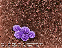
A colourised SEM of MRSA


Staphylococcus aureus (colloquially known as "Staph aureus" or a "Staph infection") is one of the major resistant pathogens. Found on the mucous membranes and the human skin of around a third of the population, it is extremely adaptable to antibiotic pressure. It was one of the earlier bacteria in which penicillin resistance was found in 1947, just four years after the drug started being mass-produced. Methicillin was then the antibiotic of choice, but has since been replaced by oxacillin due to significant kidney toxicity. Methicillin-resistant Staphylococcus aureus (MRSA) was first detected in Britain in 1961, and is now "quite common" in hospitals. MRSA was responsible for 37% of fatal cases of sepsis in the UK in 1999, up from 4% in 1991. Half of all S. aureus infections in the US are resistant to penicillin, methicillin, tetracycline and erythromycin.

This left vancomycin as the only effective agent available at the time. However, strains with intermediate (4-8 μg/ml) levels of resistance, termed glycopeptide-intermediate Staphylococcus aureus (GISA) or vancomycin-intermediate Staphylococcus aureus (VISA), began appearing in the late 1990s. The first identified case was in Japan in 1996, and strains have since been found in hospitals in England, France and the US. The first documented strain with complete (>16 μg/ml) resistance to vancomycin, termed vancomycin-resistant Staphylococcus aureus (VRSA) appeared in the United States in 2002. However, in 2011 a variant of vancomycin has been tested that binds to the lactate variation and also binds well to the original target, thus reinstates potent antimicrobial activity.

A new class of antibiotics, oxazolidinones, became available in the 1990s, and the first commercially available oxazolidinone, linezolid, is comparable to vancomycin in effectiveness against MRSA. Linezolid-resistance in S. aureus was reported in 2001.
Community-acquired MRSA (CA-MRSA) has now emerged as an epidemic that is responsible for rapidly progressive, fatal diseases, including necrotizing pneumonia, severe sepsis and necrotizing fasciitis.

MRSA is the most frequently identified antimicrobial drug-resistant pathogen in US hospitals. The epidemiology of infections caused by MRSA is rapidly changing. In the past 10 years, infections caused by this organism have emerged in the community. The two MRSA clones in the United States most closely associated with community outbreaks, USA400 (MW2 strain, ST1 lineage) and USA300, often contain Panton-Valentine leukocidin (PVL) genes and, more frequently, have been associated with skin and soft tissue infections. Outbreaks of CA-MRSA infections have been reported in correctional facilities, among athletic teams, among military recruits, in newborn nurseries, and among men who have sex with men. CA-MRSA infections now appear endemic in many urban regions and cause most CA-S. aureus infections.

2)     Streptococcus and Enterococcus:


Coloured SEM of Enterococcus faecalis bacteria


Streptococcus pyogenes (Group A Streptococcus: GAS) infections can usually be treated with many different antibiotics. Early treatment may reduce the risk of death from invasive group A streptococcal disease. However, even the best medical care does not prevent death in every case. For those with very severe illness, supportive care in an intensive care unit may be needed. For persons with necrotizing fasciitis, surgery often is needed to remove damaged tissue. Strains of S. pyogenes resistant to macrolide antibiotics have emerged; however, all strains remain uniformly sensitive to penicillin.

Resistance of Streptococcus pneumoniae to penicillin and other beta-lactams is increasing worldwide. The major mechanism of resistance involves the introduction of mutations in genes encoding penicillin-binding proteins. Selective pressure is thought to play an important role, and use of beta-lactam antibiotics has been implicated as a risk factor for infection and colonization. S. pneumoniae is responsible for pneumonia, bacteremia, otitis media, meningitis, sinusitis, peritonitis and arthritis.

Multidrug-resistant Enterococcus faecalis and Enterococcus faecium are associated with nosocomial infections. Among these strains, penicillin-resistant Enterococcus was seen in 1983, vancomycin-resistant Enterococcus in 1987, and linezolid-resistant Enterococcus in the late 1990s.

3)     Pseudomonas aeruginosa:


Pseudomonas aeruginosa is a highly prevalent opportunistic pathogen. One of the most worrisome characteristics of P. aeruginosa is its low antibiotic susceptibility, which is attributable to a concerted action of multidrug efflux pumps with chromosomally encoded antibiotic resistance genes (for example, mexAB-oprM, mexXY, etc.) and the low permeability of the bacterial cellular envelopes. Pseudomonas aeruginosa has the ability to produce HAQs and it has been found that HAQs have prooxidant effects, and overexpressing modestly increased susceptibility to antibiotics. The study experimented with the Pseudomonas aeruginosa biofilms and found that a disruption of relA and spoT genes produced an inactivation of the Stringent response (SR) in cells who were with nutrient limitation which provides cells be more susceptible to antibiotics.

4)     Clostridium difficile:


Clostridium difficile antibiotic-resistant 


Clostridium difficile is a nosocomial pathogen that causes diarrheal disease in hospitals world wide. Clindamycin-resistant C. difficile was reported as the causative agent of large outbreaks of diarrheal disease in hospitals in New York, Arizona, Florida and Massachusetts between 1989 and 1992. Geographically dispersed outbreaks of C. difficile strains resistant to fluoroquinolone antibiotics, such as ciprofloxacin and levofloxacin, were also reported in North America in 2005.

5)     Salmonella and E. coli:


Escherichia coli: proportion of invasive isolates with resistance to fluoroquinolones


Escherichia coli and Salmonella come directly from contaminated food. When both bacteria are spread, serious health conditions arise. Many people are hospitalized each year after becoming infected, with some dying as a result. By 1993, E. coli resistant to multiple fluoroquinolone variants was documented.

6)     Acinetobacter baumannii:


Pandrug-resistant Acinetobacter baumannii


On November 5, 2004, the Centers for Disease Control and Prevention (CDC) reported an increasing number of Acinetobacter baumannii bloodstream infections in patients at military medical facilities in which service members injured in the Iraq/Kuwait region during Operation Iraqi Freedom and in Afghanistan during Operation Enduring Freedom were treated. Most of these showed multidrug resistance (MRAB), with a few isolates resistant to all drugs tested.

7)     Mycobacterium tuberculosis:


Copper resistance is essential for virulence of Mycobacterium tuberculosis


Tuberculosis is increasing across the globe, especially in developing countries, over the past few years. TB resistant to antibiotics is called MDR TB (Multidrug Resistant TB). The rise of the HIV/AIDS epidemic has contributed to this.

TB was considered one of the most prevalent diseases, and did not have a cure until the discovery of Streptomycin by Selman Waksman in 1943. However, the bacteria soon developed resistance. Since then, drugs such as isoniazid and rifampin have been used. M. tuberculosis develops resistance to drugs by spontaneous mutations in its genomes. Resistance to one drug is common, and this is why treatment is usually done with more than one drug. Extensively Drug Resistant TB (XDR TB) is TB that is also resistant to the second line of drugs.

Resistance of Mycobacterium tuberculosis to isoniazid, rifampin, and other common treatments has become an increasingly relevant clinical challenge. (For more on Drug Resistant TB, visit the Multi-drug resistant tuberculosis page.)


Alternatives:

  • Prevention:

Rational use of antibiotics may reduce the chances of development of opportunistic infection by antibiotic-resistant bacteria due to dysbacteriosis.Our immune systems will cure minor bacterial infections on their own. If we give it the chance without relying on antibiotics to cure a small infection, we will be less likely to become immune or resistant to the antibiotic. It is also important to note that antibiotics will not cure viral infections. Taking an antibiotic unnecessarily to treat a viral infection can lead to the resistance of antibiotics In one study, the use of fluoroquinolones is clearly associated with Clostridium difficile infection, which is a leading cause of nosocomial diarrhea in the United States, and a major cause of death, worldwide.

Vaccines do not have the problem of resistance because a vaccine enhances the body's natural defenses, while an antibiotic operates separately from the body's normal defenses. Nevertheless, new strains may evolve that escape immunity induced by vaccines; for example an updated influenza vaccine is needed each year.

While theoretically promising, antistaphylococcal vaccines have shown limited efficacy, because of immunological variation between Staphylococcus species, and the limited duration of effectiveness of the antibodies produced. Development and testing of more effective vaccines is under way.
The Australian Commonwealth Scientific and Industrial Research Organization (CSIRO), realizing the need for the reduction of antibiotic use, has been working on two alternatives. One alternative is to prevent diseases by adding cytokines instead of antibiotics to animal feed. These proteins are made in the animal body "naturally" after a disease and are not antibiotics, so they do not contribute to the antibiotic resistance problem. Furthermore, studies on using cytokines have shown they also enhance the growth of animals like the antibiotics now used, but without the drawbacks of nontherapeutic antibiotic use. Cytokines have the potential to achieve the animal growth rates traditionally sought by the use of antibiotics without the contribution of antibiotic resistance associated with the widespread nontherapeutic uses of antibiotics currently used in the food animal production industries. Additionally, CSIRO is working on vaccines for diseases.

  • Phage therapy:

Phage therapy, an approach that has been extensively researched and used as a therapeutic agent for over 60 years, especially in the Soviet Union, represents a potentially significant but currently underdeveloped approach to the treatment of bacterial disease. Phage therapy was widely used in the United States until the discovery of antibiotics, in the early 1940s. Bacteriophages or "phages" are viruses that invade bacterial cells and, in the case of lytic phages, disrupt bacterial metabolism and cause the bacterium to lyse. Phage therapy is the therapeutic use of lytic bacteriophages to treat pathogenic bacterial infections.

Bacteriophage therapy is an important alternative to antibiotics in the current era of multidrug resistant pathogens. A review of studies that dealt with the therapeutic use of phages from 1966–1996 and few latest ongoing phage therapy projects via internet showed: phages were used topically, orally or systemically in Polish and Soviet studies. The success rate found in these studies was 80–95% with few gastrointestinal or allergic side effects. British studies also demonstrated significant efficacy of phages against Escherichia coli, Acinetobacter spp., Pseudomonas spp. and Staphylococcus aureus. US studies dealt with improving the bioavailability of phage. Phage therapy may prove as an important alternative to antibiotics for treating multidrug resistant pathogens.

Discovery of the structure of the viral protein PlyC is allowing researchers to understand the way it kills a significant range of pathogenic bacteria.


Research:

New medications:

Until recently, research and development (R&D) efforts have provided new drugs in time to treat bacteria that became resistant to older antibiotics. However: "The supply of new replacement antimicrobial agents has slowed dramatically and we face the prospect of a future where we have far fewer options in the treatment of infectious disease" The potential crisis at hand is the result of a marked decrease in industry R&D, and the increasing prevalence of resistant bacteria. Infectious disease physicians are alarmed by the prospect that effective antibiotics may not be available to treat seriously ill patients in the near future. Poor financial investment in antibiotic research has exacerbated the situation.

In research published on October 17, 2008 in Cell, a team of scientists pinpointed the place on bacteria where the antibiotic myxopyronin launches its attack, and why that attack is successful. The myxopyronin binds to and inhibits the crucial bacterial enzyme, RNA polymerase. The myxopyronin changes the structure of the switch-2 segment of the enzyme, inhibiting its function of reading and transmitting DNA code. This prevents RNA polymerase from delivering genetic information to the ribosomes, causing the bacteria to die.

In 2012, a team of the University of Leipzig modified a peptide found in honeybees. It is effective against 37 types of bacteria.
One major cause of antibiotic resistance is the increased pumping activity of microbial ABC transporters, which diminishes the effective drug concentration inside the microbial cell. ABC transporter inhibitors that can be used in combination with current antimicrobials are being tested in clinical trials and are available for therapeutic regimens.

Applications:

Antibiotic resistance is an important tool for genetic engineering. By constructing a plasmid that contains an antibiotic resistance gene as well as the gene being engineered or expressed, a researcher can ensure that when bacteria replicate, only the copies that carry the plasmid survive. This ensures that the gene being manipulated passes along when the bacteria replicates.

The most commonly used antibiotics in genetic engineering are generally "older" antibiotics that have largely fallen out of use in clinical practice.

These include:

  • ampicillin
  • kanamycin
  • tetracycline
  • chloramphenicol


Industrially the use of antibiotic resistance is disfavored since maintaining bacterial cultures would require feeding them large quantities of antibiotics. Instead, the use of auxotrophic bacterial strains (and function-replacement plasmids) is preferred.


REFERENCE:

Tuesday, June 25, 2013

Embryology


Embryology


1 - morula, 2 - blastula


1 - blastula, 2 - gastrula with blastopore; orange - ectoderm, red - endoderm


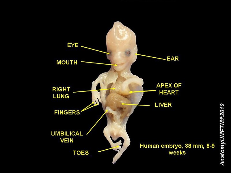
Dissection of human embryo, 38 mm - 8 weeks


Embryology  is the science that studies the development of an embryo from the ovum fertilization of the to the fetus stage.


Embryonic development of animals:

After cleavage, the dividing cells, or morula, becomes a hollow ball, or blastula, which develops a hole or pore at one end.


  • Bilaterans:

In bilateral animals, the blastula develops in one of two ways that divides the whole animal kingdom into two halves (see: Embryological origins of the mouth and anus). If in the blastula the first pore (blastopore) becomes the mouth of the animal, it is a protostome; if the first pore becomes the anus then it is a deuterostome. The protostomes include most invertebrate animals, such as insects, worms and molluscs, while the deuterostomes include the vertebrates. In due course, the blastula changes into a more differentiated structure called the gastrula.

The gastrula with its blastopore soon develops three distinct layers of cells (the germ layers) from which all the bodily organs and tissues then develop:

  1. The innermost layer, or endoderm, gives rise to the digestive organs, the gills, lungs or swim bladder if present, and kidneys or nephrites.
  2. The middle layer, or mesoderm, gives rise to the muscles, skeleton if any, and blood system.
  3. The outer layer of cells, or ectoderm, gives rise to the nervous system, including the brain, and skin or carapace and hair, bristles, or scales.

Embryos in many species often appear similar to one another in early developmental stages. The reason for this similarity is because species have a shared evolutionary history. These similarities among species are called homologous structures, which are structures that have the same or similar function and mechanism, having evolved from a common ancestor.

Humans:

Humans are bilaterans and deuterostomes - the anus develops first.
In humans, the term embryo refers to the ball of dividing cells from the moment the zygote implants itself in the uterus wall until the end of the eighth week after conception. Beyond the eighth week after conception (tenth week of pregnancy), the developing human is then called a fetus.


History of embryology:


Human embryo at six weeks gestational age


Histological film 10 day mouse embryo


Beetle larvae

As recently as the 18th century, the prevailing notion in human embryology was preformation: the idea that semen contains an embryo — a preformed, miniature infant, or "homunculus" — that simply becomes larger during development. The competing explanation of embryonic development was epigenesis, originally proposed 2,000 years earlier by Aristotle. Much early embryology came from the work of the great Italian anatomists: Aldrovandi, Aranzio, Leonardo da Vinci, Marcello Malpighi, Gabriele Falloppio, Girolamo Cardano, Emilio Parisano, Fortunio Liceti, Stefano Lorenzini, Spallanzani, Enrico Sertoli, Mauro Rusconi, etc. According to epigenesis, the form of an animal emerges gradually from a relatively formless egg. As microscopy improved during the 19th century, biologists could see that embryos took shape in a series of progressive steps, and epigenesis displaced preformation as the favoured explanation among embryologists.

  • After 1827:


8-9 weeks human embryo

Karl Ernst von Baer and Heinz Christian Pander proposed the germ layer theory of development; von Baer discovered the mammalian ovum in 1827. Modern embryological pioneers include Charles Darwin, Ernst Haeckel, J.B.S. Haldane, and Joseph Needham. Other important contributors include William Harvey, Kaspar Friedrich Wolff, Heinz Christian Pander, August Weismann, Gavin de Beer, Ernest Everett Just, and Edward B. Lewis.

  • After 1950:

After the 1950s, with the DNA helical structure being unravelled and the increasing knowledge in the field of molecular biology, developmental biology emerged as a field of study which attempts to correlate the genes with morphological change, and so tries to determine which genes are responsible for each morphological change that takes place in an embryo, and how these genes are regulated.


Vertebrate and invertebrate embryology:

Many principles of embryology apply to invertebrates as well as to vertebrates. Therefore, the study of invertebrate embryology has advanced the study of vertebrate embryology. However, there are many differences as well. For example, numerous invertebrate species release a larva before development is complete; at the end of the larval period, an animal for the first time comes to resemble an adult similar to its parent or parents. Although invertebrate embryology is similar in some ways for different invertebrate animals, there are also countless variations. For instance, while spiders proceed directly from egg to adult form many insects develop through at least one larval stage


Modern embryology research:

Currently, embryology has become an important research area for studying the genetic control of the development process (e.g. morphogens), its link to cell signalling, its importance for the study of certain diseases and mutations and in links to stem cell research.


Reference:

Sunday, June 23, 2013

What is Anatomy ?


Anatomy


The anatomy lesson of Dr. Nicolaes Tulp by Rembrandt shows an anatomy lesson taking place in Amsterdam in 1632


Michiel Jansz van MiereveltAnatomy lesson of Dr. Willem van der Meer


Anatomy is a part of biology and medicine that studies the structure of living things. Anatomy in general term includes human anatomy, animal anatomy (zootomy), and plant anatomy (phytotomy). In some of its aspects anatomy is closely related to embryology (science of the development of an embryo from the fertilization of the ovum to the fetus stage), comparative anatomy and comparative embryology, through common roots in evolution.

Anatomy is subdivided into gross anatomy (or macroscopic anatomy) and microscopic anatomy. Gross anatomy is the study of anatomical structures that can, when suitably presented or dissected, be seen by unaided vision with the naked eye. Microscopic anatomy is the study of minute anatomical structures on a microscopic scale. It includes histology (the study of tissues), and cytology (the study of cells). The terms microanatomy and histology are also sometimes used synonymously (in which case the distinction between histology and cell biology isn't strictly made as described here).

The history of anatomy has been characterized, over time, by a continually developing understanding of the functions of organs and structures in the body. Methods have also improved dramatically, advancing from examination of animals through dissection of cadavers (dead human bodies) to technologically complex techniques developed in the 20th century including X-ray, ultrasound, and MRI.


Superficial anatomy:

Superficial anatomy or surface anatomy is important in anatomy being the study of anatomical landmarks that can be readily seen from the contours or the surface of the body. With knowledge of superficial anatomy, physicians or veterinary surgeons gauge the position and anatomy of the associated deeper structures. Superficial is a directional term that indicates one structure is located more externally than another, or closer to the surface of the body.


Animal anatomy:

  • Insect anatomy:


Insect morphology

Legend of body parts
Tagmata : A - Head, B - Thorax, C - Abdomen.

1. antenna
17. anus
2. ocelli (lower)
18. oviduct
3. ocelli (upper)
19. nerve chord (abdominal ganglia)
4. compound eye
20. Malpighian tubes
5. brain (cerebral ganglia)
21. tarsal pads
6. prothorax
22. claws
7. dorsal blood vessel
23. tarsus
8. tracheal tubes (trunk with spiracle)
24. tibia
9. mesothorax
25. femur
10. metathorax
26. trochanter
11. forewing
27. fore-gut (crop, gizzard)
12. hindwing
28. thoracic ganglion
13. mid-gut (stomach)
29. coxa
14. dorsal tube (Heart)
30. salivary gland
15. ovary
31. subesophageal ganglion
16. hind-gut (intestine, rectum & anus)
32. mouthparts


Insects possess segmented bodies supported by an exoskeleton, a hard-jointed outer covering made mostly of chitin. The segments of the body are organized into three distinct parts, a head, a thorax and an abdomen. The head typically bears a pair of sensory antennae, a pair of compound eyes, one to three simple eyes (ocelli) and three sets of modified appendages that form the mouthparts. The thorax has three pairs of segmented legs, one pair each for the three segments that compose the thorax and one or two pairs of wings. The abdomen is composed of eleven segments, some of which may be fused and houses the digestive, respiratory, excretory and reproductive systems. There is considerable variations between species and many adaptations to the body parts, especially wings, legs, antennae and mouthparts.


  • Vertebrate anatomy:


Fossilized skeleton of Diplodocus carnegii, showing an extreme example of the backbone that characterizes the vertebrates. Exhibited at the Museum für Naturkunde (Museum of Natural Science), Berlin.


Vertebrates include the overwhelming majority of the phylum Chordata, with currently about 64,000 species described. Vertebrates include the jawless fish, bony fish, sharks and rays, amphibians, reptiles, mammals, and birds. Extant vertebrates range in size from the frog species Paedophryne amauensis, at as little as 7.7 mm (0.3 inch), to the blue whale, at up to 33 m (110 ft). Vertebrates make up about 4% of all described animal species; the rest are invertebrates, which lack backbones.

1)     Bird anatomy:


External anatomy (topography) of a typical bird: 1 Beak, 2 Head, 3 Iris, 4 Pupil, 5 Mantle, 6 Lesser coverts, 7 Scapulars, 8 Coverts, 9 Tertials, 10 Rump, 11 Primaries, 12 Vent, 13 Thigh, 14 Tibio-tarsal articulation, 15 Tarsus, 16 Feet, 17 Tibia, 18 Belly, 19 Flanks, 20 Breast, 21 Throat, 22 Wattle, 23 Eyestripe


Birds are quadrupeds but though their hind limbs are used for walking or hopping, their front limbs are wings covered with feathers and adapted for flight. Birds are endothermic, have a high metabolic rate, a light skeletal system and powerful muscles. The long bones are thin, hollow and very light. Air sac extensions from the lungs occupy the centre of some bones. The sternum is wide and usually has a keel and the caudal vertebrae are fused. There are no teeth and the narrow jaws are adapted into a horn-covered beak. The eyes are relatively large, particularly in nocturnal species such as owls. They face forwards in predators and sideways in ducks.

The feathers are outgrowths of the epidermis. Large flight feathers are found on the wing and tail, contour feathers cover the bird's surface and fine down occurs on young birds and under the contour feathers of water birds. There are scales on the legs and feet and claws on the tips of the toes.

2)     Reptile anatomy:


Clockwise from above left: Green turtle (Chelonia mydas), tuatara (Sphenodon punctatus), Nile crocodile (Crocodylus niloticus), and Sinai agama (Pseudotrapelus sinaitus)


Traditionally, reptiles are members of the class Reptilia comprising the amniotes that are neither birds nor mammals. (The amniotes are the vertebrates with eggs featuring an amnion, a double membrane that permits the embryo to breathe effectively on land.) Living reptiles, in that sense, can be distinguished from other tetrapods in that they are "cold-blooded" (poikilothermic) and bear scutes or scales.

3)     Fish anatomy:


Skeleton of a butterfly fish showing the vertebral column and fin rays


The body of a fish is divided into a head, trunk and tail, although the divisions between the three are not always externally visible. The skeleton, which forms the support structure inside the fish, is either made of cartilage (cartilaginous fish) or bone (bony fishes). The main skeletal element is the vertebral column, composed of articulating vertebrae which are lightweight yet strong. The ribs attach to the spine and there are no limbs or limb girdles. The main external features of the fish, the fins, are composed of bony spines and soft rays and, with the exception of the caudal fins, have no direct connection with the spine. They are supported by the muscles which compose the main part of the trunk.

4)     Mammal anatomy:


Examples of various mammalian orders, click the image and scroll down for individual descriptions


Mammals are a clade of endothermic amniotes. Among the features that distinguish them from the other amniotes, the reptiles and the birds, are hair, three middle ear bones, mammary glands in females, and a neocortex (a region of the brain). The mammalian brain regulates body temperature and the circulatory system, including the four-chambered heart. The mammals include the largest animals on the planet, the rorqual whales, as well as some of the most intelligent, such as elephants, some primates and some cetaceans. The basic body type is a four-legged land-borne animal, but some mammals are adapted for life at sea, in the air, in the trees, or on two legs. The largest group of mammals, the placentals, have a placenta which feeds the offspring during pregnancy. Mammals range in size from the 30–40 mm (1.2–1.6 in) bumblebee bat to the 33-meter (108 ft) blue whale.

  • Human anatomy:


Para-sagittal MRI scan of the head


An X-ray of a human chest


Human heart and lungs, from an old edition of Gray's Anatomy


Human anatomy, including gross human anatomy and histology, is primarily the scientific study of the morphology of the adult human body. It differs from Physiology in that Anatomy is only the structures involved, and Physiology is the way those structures actually work.

Generally, students of certain biological sciences, paramedics, prosthetists and orthotists, physiotherapists, occupational therapists, nurses, and medical students learn gross anatomy and microscopic anatomy from anatomical models, skeletons, textbooks, diagrams, photographs, lectures and tutorials, and in addition, medical students generally also learn gross anatomy through practical experience of dissection and inspection of cadavers. The study of microscopic anatomy (or histology) can be aided by practical experience examining histological preparations (or slides) under a microscope.

Human anatomy, physiology and biochemistry are complementary basic medical sciences, which are generally taught to medical students in their first year at medical school. Human anatomy can be taught regionally or systemically; that is, respectively, studying anatomy by bodily regions such as the head and chest, or studying by specific systems, such as the nervous or respiratory systems. The major anatomy textbook, Gray's Anatomy, has recently been reorganized from a systems format to a regional format, in line with modern teaching methods. A thorough working knowledge of anatomy is required by physicians, especially surgeons and doctors working in some diagnostic specialties, such as histopathology and radiology.

Academic human anatomists are usually employed by universities, medical schools or teaching hospitals. They are often involved in teaching anatomy, and research into certain systems, organs, tissues or cells.


Plant anatomy:

Plant anatomy involves the structure of the cells, in contrast to animal cells. Plant cells have cell walls, chlorophyll, and lack mitochondria. They also contain larger reservoirs called Lysosomes. Plant lack the digestive organs common among animals because they create their own energy from the sun, in photosynthesis. This makes them more independent and also a source of energy for other animals.


Other branches:


  • Comparative anatomy relates to the comparison of anatomical structures (both gross and microscopic) in different animals.
  • Anthropological anatomy or physical anthropology relates to the comparison of the anatomy of different races of humans.
  • Artistic anatomy relates to anatomic studies for artistic reasons.


Saturday, June 22, 2013

Analytical chemistry


Analytical chemistry


Analytical chemistry is the study of the chemical components of artificial and natural materials through identification, quantification, and separation.

  • Qualitative analysis determines the indication of the identity of the chemical species in the sample.
  • Quantitative analysis determines the amount of certain components in the substancein the sample.
  • The separation of components is often performed before starting of the analysis.

Analytical methods can be divided into classical and instrumental.
Classical methods (also known as wet chemistry methods) use separations such as:

  1. precipitation
  2. extraction
  3. distillation
  4. qualitative analysis by color, odor, or melting point.
Quantitative analysis is achieved by measurement of weight or volume

Instrumental methods use an apparatus to measure physical quantities of the analyte such as:

  1. light absorption
  2. fluorescence
  3. conductivity

The separation of materials is accomplished using chromatography, electrophoresis or Field Flow Fractionation methods.

Analytical chemistry is also focused on improvements in experimental design, chemometrics, and the creation of new measurement tools to provide better chemical information. Analytical chemistry has applications in forensics, bioanalysis, clinical analysis, environmental analysis, and materials analysis.


History of Analytical chemistry:


Gustav Kirchhoff (left) and Robert Bunsen (right)

Analytical chemistry has been important since the early days of chemistry, providing methods for determining which elements and chemicals are present in the object in question. During this period significant analytical contributions to chemistry include the development of systematic elemental analysis by Justus von Liebig and systematized organic analysis based on the specific reactions of functional groups.
The first instrumental analysis was flame emissive spectrometry developed by Robert Bunsen and Gustav Kirchhoff who discovered rubidium (Rb) and caesium (Cs) in 1860.

Most of the major developments in analytical chemistry take place after 1900. During this period instrumental analysis becomes progressively dominant in the field. In particular many of the basic spectroscopic and spectrometric techniques were discovered in the early 20th century and refined in the late 20th century.
The separation sciences follow a similar time line of development and also become increasingly transformed into high performance instruments. In the 1970s many of these techniques began to be used together to achieve a complete characterization of samples.

Starting in approximately the 1970s into the present day analytical chemistry has progressively become more inclusive of biological questions (bioanalytical chemistry), whereas it had previously been largely focused on inorganic or small organic molecules. Lasers have been increasingly used in chemistry as probes and even to start and influence a wide variety of reactions. The late 20th century also saw an expansion of the application of analytical chemistry from somewhat academic chemical questions to forensic, environmental, industrial and medical questions, such as in histology.

Modern analytical chemistry is dominated by instrumental analysis. Many analytical chemists focus on a single type of instrument. Academics tend to either focus on new applications and discoveries or on new methods of analysis. The discovery of a chemical present in blood that increases the risk of cancer would be a discovery that an analytical chemist might be involved in. An effort to develop a new method might involve the use of a tunable laser to increase the specificity and sensitivity of a spectrometric method. Many methods, once developed, are kept purposely static so that data can be compared over long periods of time. This is particularly true in industrial quality assurance (QA), forensic and environmental applications. Analytical chemistry plays an increasingly important role in the pharmaceutical industry where, aside from QA, it is used in discovery of new drug candidates and in clinical applications where understanding the interactions between the drug and the patient are critical.


Classical methods:


The presence of copper in this qualitative analysis is indicated by the bluish-green color of the flame

Although modern analytical chemistry is dominated by sophisticated instrumentation, the roots of analytical chemistry and some of the principles used in modern instruments are from traditional techniques many of which are still used today. These techniques also tend to form the backbone of most undergraduate analytical chemistry educational labs.


Qualitative analysis:

A qualitative analysis determines the presence or absence of a particular compound, but not the mass or concentration. That is, if it is not related to quantity.


  • Chemical tests:


There are numerous qualitative chemical tests, for example, the acid test for gold and the Kastle-Meyer test for the presence of blood.


  • Flame test:

Inorganic qualitative analysis generally refers to a systematic scheme to confirm the presence of certain, usually aqueous, ions or elements by performing a series of reactions that eliminate ranges of possibilities and then confirms suspected ions with a confirming test. Sometimes small carbon containing ions are included in such schemes. With modern instrumentation these tests are rarely used but can be useful for educational purposes and in field work or other situations where access to state-of-the-art instruments are not available or expedient.


Gravimetric analysis:

Gravimetric analysis involves determining the amount of material present by weighing the sample before and/or after some transformation. A common example used in undergraduate education is the determination of the amount of water in a hydrate by heating the sample to remove the water such that the difference in weight is due to the loss of water.


Volumetric analysis:

Titration involves the addition of a reactant to a solution being analyzed until some equivalence point is reached. Often the amount of material in the solution being analyzed may be determined. Most familiar to those who have taken chemistry during secondary education is the acid-base titration involving a color changing indicator. There are many other types of titrations, for example potentiometric titrations. These titrations may use different types of indicators to reach some equivalence point.


Instrumental methods:


Block diagram of an analytical instrument showing the stimulus and measurement of response


  • Spectroscopy:

Spectroscopy measures the interaction of the molecules with electromagnetic radiation. Spectroscopy consists of many different applications such as atomic absorption spectroscopy, atomic emission spectroscopy, ultraviolet-visible spectroscopy, x-ray fluorescence spectroscopy, infrared spectroscopy, Raman spectroscopy, dual polarisation interferometry, nuclear magnetic resonance spectroscopy, photoemission spectroscopy, Mössbauer spectroscopy and so on.


  • Mass spectrometry:


An accelerator mass spectrometer used for radiocarbon dating and other analysis

Mass spectrometry measures mass-to-charge ratio of molecules using electric and magnetic fields. There are several ionization methods: electron impact, chemical ionization, electrospray, fast atom bombardment, matrix assisted laser desorption ionization, and others. Also, mass spectrometry is categorized by approaches of mass analyzers: magnetic-sector, quadrupole mass analyzer, quadrupole ion trap, time-of-flight, Fourier transform ion cyclotron resonance, and so on.


  • Electrochemical analysis:

Electroanalytical methods measure the potential (volts) and/or current (amps) in an electrochemical cell containing the analyte. These methods can be categorized according to which aspects of the cell are controlled and which are measured. The three main categories are potentiometry (the difference in electrode potentials is measured), coulometry (the cell's current is measured over time), and voltammetry (the cell's current is measured while actively altering the cell's potential).

  • Thermal analysis:

Further information: Calorimetry, thermal analysis
Calorimetry and thermogravimetric analysis measure the interaction of a material and heat.

  • Separation:


Separation of black ink on a thin layer chromatography plate

Further information: Separation process, Chromatography, electrophoresis
Separation processes are used to decrease the complexity of material mixtures. Chromatography, electrophoresis and Field Flow Fractionation are representative of this field.

  • Hybrid techniques:

Combinations of the above techniques produce a "hybrid" or "hyphenated" technique.
Several examples are in popular use today and new hybrid techniques are under development. For example, gas chromatography-mass spectrometry, gas chromatography-infrared spectroscopy, liquid chromatography-mass spectrometry, liquid chromatography-NMR spectroscopy. liquid chromagraphy-infrared spectroscopy and capillary electrophoresis-mass spectrometry.

Hyphenated separation techniques refers to a combination of two (or more) techniques to detect and separate chemicals from solutions. Most often the other technique is some form of chromatography. Hyphenated techniques are widely used in chemistry and biochemistry. A slash is sometimes used instead of hyphen, especially if the name of one of the methods contains a hyphen itself.

  • Microscopy:


Fluorescence microscope image of two mouse cell nuclei in prophase (scale bar is 5 µm)

The visualization of single molecules, single cells, biological tissues and nanomaterials is an important and attractive approach in analytical science. Also, hybridization with other traditional analytical tools is revolutionizing analytical science. Microscopy can be categorized into three different fields: optical microscopy, electron microscopy, and scanning probe microscopy. Recently, this field is rapidly progressing because of the rapid development of the computer and camera industries.

  • Lab-on-a-chip:


A glass microreactor

Devices that integrate (multiple) laboratory functions on a single chip of only millimeters to a few square centimeters in size and that are capable of handling extremely small fluid volumes down to less than pico liters.


Standards:

  • Standard curve:


A calibration curve plot showing limit of detection (LOD), limit of quantification (LOQ), dynamic range, and limit of linearity (LOL)

A general method for analysis of concentration involves the creation of a calibration curve. This allows for determination of the amount of a chemical in a material by comparing the results of unknown sample to those of a series known standards. If the concentration of element or compound in a sample is too high for the detection range of the technique, it can simply be diluted in a pure solvent. If the amount in the sample is below an instrument's range of measurement, the method of addition can be used. In this method a known quantity of the element or compound under study is added, and the difference between the concentration added, and the concentration observed is the amount actually in the sample.

  • Internal standards:

Sometimes an internal standard is added at a known concentration directly to an analytical sample to aid in quantitation. The amount of analyte present is then determined relative to the internal standard as a calibrant. An ideal internal standard is isotopically-enriched analyte which gives rise to the method of isotope dilution.

  • Standard addition:

The method of standard addition is used in instrumental analysis to determine concentration of a substance (analyte) in an unknown sample by comparison to a set of samples of known concentration, similar to using a calibration curve. Standard addition can be applied to most analytical techniques and is used instead of a calibration curve to solve the matrix effect problem.


Signals and noise:

One of the most important components of analytical chemistry is maximizing the desired signal while minimizing the associated noise. The analytical figure of merit is known as the signal-to-noise ratio (S/N or SNR)
Noise can arise from environmental factors as well as from fundamental physical processes.

  • Thermal noise:

Thermal noise results from the motion of charge carriers (usually electrons) in an electrical circuit generated by their thermal motion. Thermal noise is white noise meaning that the power spectral density is constant throughout the frequency spectrum.

The root mean square value of the thermal noise in a resistor is given by:


where
  kB is Boltzmann's constant
  T is the temperature
  R is the resistance
is the bandwidth of the frequency f

  • Shot noise:

Shot noise is a type of electronic noise that occurs when the finite number of particles (such as electrons in an electronic circuit or photons in an optical device) is small enough to give rise to statistical fluctuations in a signal.
Shot noise is a Poisson process and the charge carriers that make up the current follow a Poisson distribution.

The root mean square current fluctuation is given by:


where e is the elementary charge and I is the average current. Shot noise is white noise.

  • Flicker noise:

Flicker noise is electronic noise with a 1/ƒ frequency spectrum; as f increases, the noise decreases. Flicker noise arises from a variety of sources, such as impurities in a conductive channel, generation and recombination noise in a transistor due to base current, and so on. This noise can be avoided by modulation of the signal at a higher frequency, for example through the use of a lock-in amplifier.

  • Environmental noise:


Noise in a thermogravimetric analysis; lower noise in the middle of the plot results from less human activity (and environmental noise) at night

Environmental noise arises from the surroundings of the analytical instrument. Sources of electromagnetic noise are power lines, radio and television stations, wireless devices, Compact fluorescent lamps and electric motors. Many of these noise sources are narrow bandwidth and therefore can be avoided. Temperature and vibration isolation may be required for some instruments.

  • Noise reduction:

Noise reduction can be accomplished either in computer hardware or software. Examples of hardware noise reduction are the use of shielded cable, analog filtering, and signal modulation. Examples of software noise reduction are digital filtering, ensemble average, boxcar average, and correlation methods.


Applications:


Analytical chemistry research is largely driven by performance (sensitivity, selectivity, robustness, linear range, accuracy, precision, and speed), and cost (purchase, operation, training, time, and space). Among the main branches of contemporary analytical atomic spectrometry, the most widespread and universal are optical and mass spectrometry.

In the direct elemental analysis of solid samples, the new leaders are laser-induced breakdown and laser ablation mass spectrometry, and the related techniques with transfer of the laser ablation products into inductively coupled plasma. Advances in design of diode lasers and optical parametric oscillators promote developments in fluorescence and ionization spectrometry and also in absorption techniques where uses of optical cavities for increased effective absorption pathlength are expected to expand. The use of plasma- and laser-based methods is increasing. An interest towards absolute (standardless) analysis has revived, particularly in emission spectrometry.

great effort is put in shrinking the analysis techniques to chip size. Although there are few examples of such systems competitive with traditional analysis techniques, potential advantages include size/portability, speed, and cost. (micro Total Analysis System (µTAS) or Lab-on-a-chip). Microscale chemistry reduces the amounts of chemicals used.

Many development improve the analysis of biological systems. Examples of rapidly expanding fields in this area are:

  • Genomics - DNA sequencing and its related research. Genetic fingerprinting and DNA microarray are important tools and research fields.
  • Proteomics - the analysis of protein concentrations and modifications, especially in response to various stressors, at various developmental stages, or in various parts of the body.
  • Metabolomics - similar to proteomics, but dealing with metabolites.
  • Transcriptomics - mRNA and its associated field
  • Lipidomics - lipids and its associated field
  • Peptidomics - peptides and its associated field
  • Metalomics - similar to proteomics and metabolomics, but dealing with metal concentrations and especially with their binding to proteins and other molecules.

Analytical chemistry has played critical roles in the understanding of basic science to a variety of practical applications, such as biomedical applications, environmental monitoring, quality control of industrial manufacturing, forensic science and so on.

The recent developments of computer automation and information technologies have extended analytical chemistry into a number of new biological fields. For example, automated DNA sequencing machines were the basis to complete human genome projects leading to the birth of genomics. Protein identification and peptide sequencing by mass spectrometry opened a new field of proteomics.

Analytical chemistry has been an indispensable area in the development of nanotechnology. Surface characterization instruments, electron microscopes and scanning probe microscopes enables scientists to visualize atomic structures with chemical characterizations.